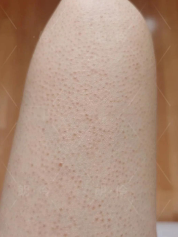
You’re on vacation, or perhaps just spending more time in the summer sun, when you notice something odd. While the skin on your left thigh tans (or burns) evenly, there’s one distinct patch—perhaps the size of a palm or larger—that remains stubbornly, starkly pale. It doesn’t darken. It doesn’t redden. It’s like a permanent island of winter skin amidst a summer sea. This isn’t a scar or a recent sunblock miss. This persistent, non-reactive patch is far more interesting. It is a genetic marker, a visible flag planted by your own DNA, and it can be a potential indicator for two significant things: 1) A localized somatic mutation, or 2) An underlying neurocutaneous condition.
This phenomenon is a dramatic lesson in developmental biology, written on your skin.
The Genetic Blueprint: Your Personal Mosaic
Every one of us is a genetic mosaic. Very early in embryonic development, as cells divided to form your skin, a single cell may have undergone a spontaneous mutation in a gene critical for pigment production. All the millions of skin cells that descended from that one mutant cell will carry that same mutation. They will grow to form a distinct patch of skin with different properties than the surrounding “normal” cells. This patch is called a Blaschkoid patch or, more broadly, a manifestation of mosaicism.
The most common and benign form of this is Nevus Depigmentosus (also called achromic nevus). This is a stable, well-defined pale patch present from birth or early childhood where melanocytes (pigment cells) are present but produce less melanin. It’s your personal, harmless genetic signature.
The Potential Neurological Link: When Skin and Nerves Share a Blueprint
Here is where the signal becomes more significant. The skin and the nervous system develop from the same embryonic layer (the ectoderm). A visible anomaly in the skin can sometimes be a clue to a parallel, invisible anomaly in the underlying nerve tissue. This is the core concept behind neurocutaneous syndromes.
The pale patch that never tans could be a cutaneous marker for:
- Nevus Depigmentosus as a Minor Form of Mosaicism: While usually isolated, extensive or multiple patches can rarely be associated with mosaic disorders that might affect other systems. A doctor would look for subtle asymmetries in limb size or muscle tone.
- A Subtle Sign of Mosaic Neurofibromatosis Type 1 (NF1): While the classic café-au-lait spots of NF1 are dark, individuals with mosaic or segmental NF1 (where the mutation is not in all cells) can sometimes present with hypopigmented (lighter) patches as their primary skin sign. These patches follow Blaschko’s lines (the invisible developmental pathways of skin cells). Under a Wood’s lamp, they may glow more brightly.
- Hypomelanosis of Ito: This is a neurocutaneous syndrome characterized by whorled, streaked, or patchy hypopigmentation (exactly like your pale patch) following Blaschko’s lines. Crucially, it is associated with a high incidence of neurological issues (seizures, intellectual disability), musculoskeletal abnormalities, and eye problems. The skin finding is the primary visible clue.
The “Potential” It Signals: A Window into Your Wiring
Therefore, a congenital, non-reactive pale patch is a genetic marker for potential:
- Localized Genetic Variance: A fascinating quirk of your personal development with no health implications.
- Underlying Mosaicism with Systemic Effects: The possibility that the same early developmental mutation that affected that skin patch could also be present in other cell lines, potentially affecting nerve, bone, or muscle development in that same segment of the body.
Your Action Plan: From Curiosity to Clarity
- Document Its History. Has it been there since childhood? Has it changed in size or shape? (Stable is reassuring; changing is not).
- Examine Its Borders and Pattern. Does it look like a random splash, or does it have a swirling, linear, or “splash of paint” pattern? Swirling patterns that follow Blaschko’s lines are more linked to genetic mosaicism.
- Perform the Wood’s Lamp Test. In a dark room, shine a Wood’s lamp (a black light, available online) on the patch. If it accentuates the contrast, making it glow a bright, chalky white, it suggests a true absence of pigment and is more significant than simple pallor.
- Conduct a Full-Body Skin Check. Are there any other unusual patches, light or dark? Any soft, fleshy lumps (neurofibromas)? Any asymmetry in your limbs?
- See a Dermatologist, Preferably One Specializing in Genetic Skin Disorders. This is key. Show them the patch. Describe its lifelong stability. They can make a clinical diagnosis.
- If the Pattern is Extensive or Whorled, a Neurological Consultation May Be Advised. This is not to alarm, but to be thorough. A neurologist might ask about seizures, learning difficulties, or muscle weakness, and may recommend a baseline MRI if the skin findings are extensive and suggestive of a syndrome like Hypomelanosis of Ito.
That lone, pale patch on your thigh is more than a spot that missed the sun. It is a fossilized map of your earliest cellular journey. It marks a clone of cells that have been different since the beginning, marching to the beat of a slightly different genetic drummer. By understanding its origin, you transform it from a curiosity into a key—a key that can, in certain patterns, unlock important information about your unique developmental blueprint and guide you toward proactive, informed healthcare. It is the silent, visible whisper of your embryonic past.